

1.5T High-field MRI
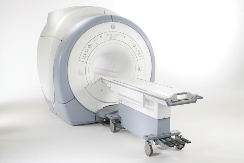
Carlsbad Imaging offers the Signa HDxt 1.5T High-field MRI system from GE Healthcare and combines advanced applications and workflow improvements with the high definition platform you know and trust. Features include: A foundation of high-performance components, whose integration enables superior imaging capabilities that aim to bring you the clearest, crispest images possible.
The Signa HDxt 1.5T High-field MRI is engineered for enhanced image contrast, reduced blurring and reduced artifacts so your doctor can see more. This system also incorporates motion correction to help reduce the need for rescans.
Imperial Radiology has the GE EchoSpeed 1.5 MRI scanner. This scanner is a high resolution, whole body imaging system known for its excellent performance. The powerful magnetic resonance imaging platform provides high definition results and a comprehensive range of applications.
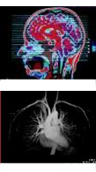
Clinical Applications
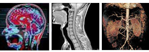 Breast
- Head and spine
- Vascular including brain, carotids, chest, abdomen and extremities
- MRCP and MR bowel
- MR myelography
- All joints including hip, shoulder, knee, elbow, ankle, foot, wrist and hand
Open MRI
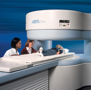 The mid-field Hitachi Open MRI unit, with its advanced gradient coil technology compares favorably with the high-field MRI machine in obtaining high-resolution images of the brain, spine and musculoskeletal anatomy. It is also excellent in acquiring MR angiographic images.
The Open unit, in addition to its fine imaging capabilities, features amazingly easy access for the patient with plenty of breathing space.
The innovative gantry design and wide patient table delivers a high level of patient comfort.
The Hitachi Open MRI offers more room than any other open MRI and is ideal for anxious patients and patients weighing up to 500 pounds.
What does MRI do for you and your medical team?
Magnetic Resonance Imaging (MRI) is one form of imaging modality used by physicians to look inside the human body to obtain clinically useful diagnostic information. Incorporating an advanced technology, MRI produces images of the anatomy without the use of radiation required with other imaging modalities such as x-ray and CT scanning.
MRI combines the physical properties of strong magnetic fields with radio waves to produce computer generated soft tissue images within any plane of the body. This widely used imaging technique can be used as a primary diagnostic tool to provide a quick and accurate diagnosis for your physician. In some situations, this procedure can reduce the need for further diagnostic procedures or invasive procedures such as exploratory surgery that may have associated complications.
What are the benefits vs. risks of MRI?
MRI is a non-invasive procedure with no known side effects. The procedure is painless. A knocking sound will be heard, which is simply the imaging process in operation.
The benefits of magnetic resonance imaging are many, with new applications continually being developed through on-going research. The procedure is used for all parts of the body and is effective in the clinical evaluation of the following conditions:
- Brain disorders
- Traumatic injuries
- Eye abnormalities
- Spine diseases
- Tumor detection
- Liver and other abdominal diseases
- Knee and shoulder injuries
- Musculoskeletal disorders
- Facial/neck abnormalities
- Infection
- Cardiac malformations
- Blood flow and vessel disorders
How to prepare for MRI?
No special preparation is required prior to the MRI exam. You may eat normally and go about your daily routine. Continue to take any medication prescribed by your doctor unless otherwise directed.
Prior to entering the scan room for your exam, you will be asked to leave those items that are not compatible with a magnetic field in a safe place outside the scan room. A list of some of these items is listed below.
- Coins
- Jewelry
- Watches
- Glasses
- Credit cards
- Hearing aids
- Keys
- Hair pins
- Other metal objects
You may also be asked to remove dentures and to wear a hospital gown to avoid magnetic interference from belt buckles and zippers.
Once you are situated on the table, make sure you are comfortable so that it is easy to remain still for the duration of the examination. Breathe normally. Once the examination has begun, you will hear a knocking sound that represents changes in the magnetic field that are a normal part of the imaging process. At the conclusion of the exam, the technologist will assist you out of the scan room.
As mentioned previously, you will be asked to leave items that are incompatible with the magnetic field outside of the scan room. Other items to consider are the presence of implants and similar items. Check with your physician or MRI technologist if you have had any brain, ear, eye or other surgeries or any of the following:
- Pacemaker
- Neuro-stimulator (Tens-unit)
- Metal implants
- Intrauterine device (IUD), etc.
- Aneurysm clips
- Surgical staples
- Implanted drug infusion device
- Foreign metal objects in the eye
- Shrapnel or bullet wounds
- Permanent eyeliner
Additional prep instructions: Note- Your specific prep instructions will be explained to you at the time your appointment is confirmed.
What happens during the MRI procedure?
A typical procedure averages 30 minutes or longer depending on the type of information required by your physician. You can help to make your images spectacular by simply relaxing and remaining as still as possible during the exam. In fact, some patients fall asleep during the MRI exam.
During your MRI examination, a technologist will be with you and will be able to see you at all times. For your convenience, an intercom system is built into the MR imager so that if you need anything, the technologist will be right there.
In certain instances, a contrast agent may be administered to enhance the study. There are no extra precautions when contrast is used.