

Introducing the new Philips Affiniti Ultrasound at Carlsbad Imaging Center
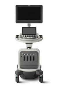 The new Philips Affiniti ultrasound system delivers the right balance of elegance and precision engineering.
Key features include:
- Crisp, clear images that enable fast, confident diagnosis and reduce the need for additional exams.
- Philips’ PureWave transducer technology, delivering excellent image quality with little or no need for image adjustment for technically difficult patients.
- Anatomical Intelligent Ultrasound, providing automatic anatomy recognition and quantification, making it easy to perform exams and quickly deliver new levels of clinical information. It is the only system in its class with PureWave Imaging which features a wide dynamic range to deliver superb spatial and contrast resolution, outstanding tissue uniformity, fewer artifacts and reduced image clutter.
- Outstanding image quality combines with advanced clinical functionality, including elastography, contrast-enhanced ultrasound (CEUS), and Anatomical Intelligence Ultrasound (AIUS).
Ultrasound is a useful way of examining many of the body’s internal organs, including but not limited to the heart, liver, gallbladder, spleen, pancreas, kidneys, and bladder.
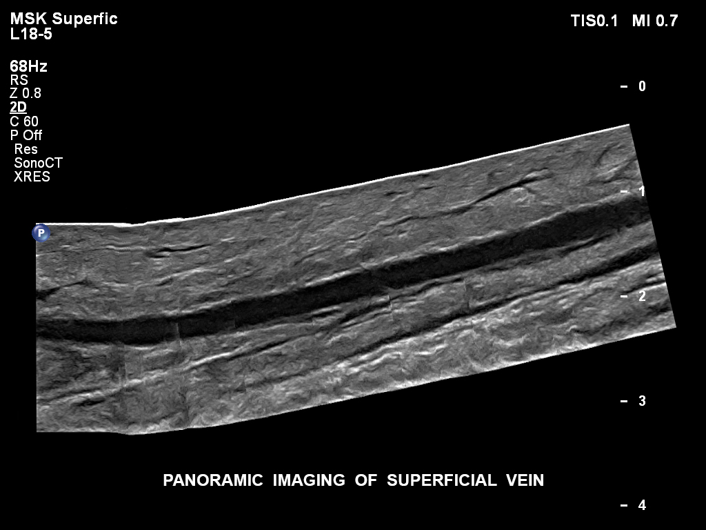
Carlsbad Imaging and Imperial Radiology offer advanced ultrasound technology. This high-resolution scanning can be performed, not only of the fetus, but of the abdominal and pelvic organs, and also of the vascular and musculoskeletal systems. It is used for the most delicate conditions without major side effects and provides extremely detailed images.
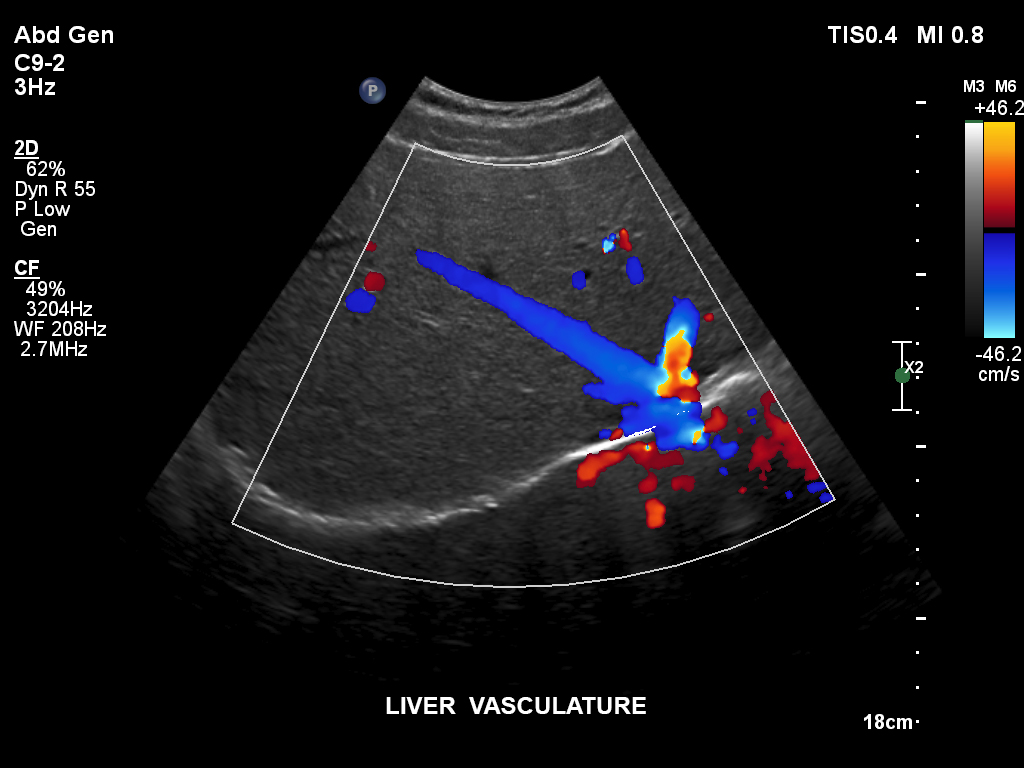
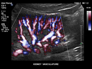
With our color Doppler imaging capabilities, we offer extremely accurate anatomic visualization, as well as valuable physiologic information of the arteries and veins in the neck, abdomen, legs and arms. Ultrasound is non-invasive, allowing physicians to diagnose without entering the body with dyes, radiation or exploratory surgery.
What is General Ultrasound Imaging?
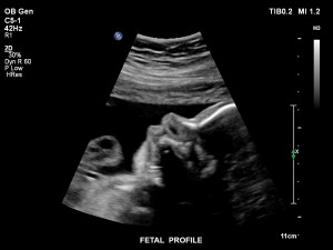
Ultrasound imaging, also called ultrasound scanning or sonography, is a method of obtaining images from inside the human body through the use of high-frequency sound waves. The reflected sound wave echoes are recorded and displayed as a real-time visual image. No ionizing radiation (x-ray) is involved in ultrasound imaging. Obstetric ultrasound refers to the specialized use of sound waves to visualize and thus determine the condition of a pregnant woman and her embryo or fetus.
Ultrasound is a useful way of examining many of the body’s internal organs, including but not limited to the heart, liver, gallbladder, spleen, pancreas, kidneys, and bladder. Because ultrasound images are captured in real time, they can show movement of internal tissues and organs and enable physicians to see blood flow.
Ultrasound imaging is based on the same principles involved in the sonar used by bats, ships at sea and anglers with fish detectors. As the sound passes through the body, echoes are produced that can be used to identify how far away an object is, how large it is, its shape and its consistency (fluid, solid or mixed).
What are the benefits vs. risks of ultrasound?
Benefits
- Ultrasound scanning is noninvasive (no needles or injections) and is usually painless.
- Ultrasound is widely available and easy to use.
- Ultrasound uses no ionizing radiation and is the preferred image modality for diagnosis and monitoring of pregnant women and their unborn infants.
- Ultrasound images can visualize structure, movement and live function in the body’s organs and blood vessels.
Risks
For standard diagnostic ultrasound there are no known harmful effects on humans.
How to prepare for ultrasound?
You should wear comfortable, loose-fitting clothing for your ultrasound exam. Other preparation depends on the type of examination you will have. For some scans, such as abdominal exam, the center may instruct you not to eat or drink for 6-8 hours before your appointment. For other scans, such as pelvic exam, you may be asked to drink up to 6 glasses of water two hours prior to your exam and avoid urinating so that your bladder is full when the scan begins.
Abdomen: No food or liquids for 8 hours prior to your exam. Take your medication as usual, a little bit of water is ok.
Renal: Full bladder required. Drink up to 6 glasses and do not urinate prior to the study.
Pelvic Ultrasound: Full bladder required. Drink up to 6 glasses and do not urinate prior to the study.
What happens during the ultrasound procedure?
Most ultrasound examinations are painless, fast and easy. You will lie on your back on an examining table. The technologist will spread gel on your skin and then press the transducer against your body, moving it until the desired images are captured. The examination usually takes less than 30 minutes.